

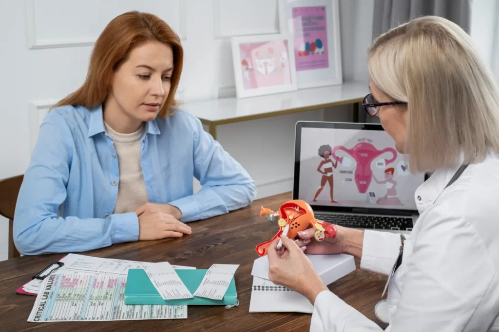
When navigating reproductive health, the true architecture of natural conception often relies entirely on a pair of sophisticated, yet highly vulnerable, biological pathways. Understanding where are the fallopian tubes located and how they function is a crucial piece of the fertility puzzle. Even a minor disruption within this delicate environment can completely alter your pregnancy and fertility trajectory, making it a vital area of focus in modern medicine.
Despite how frequently they are mentioned, many individuals still find themselves searching for answers to basic questions, such as exactly where is fallopian tube tissue positioned. Uncovering the intricate mechanics of the fallopian tube is the first step toward proactive health management. Let’s explore why fallopian tubes play such a decisive role in the body and how to recognize the hidden signs of structural distress.
What Is a Fallopian Tube?
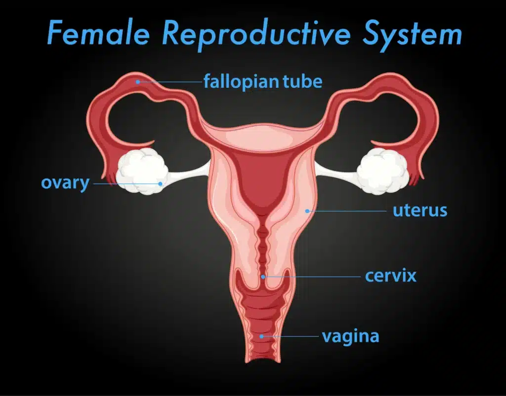
To understand how natural conception occurs, one must look at the specific anatomy of the female pelvis. Structurally, what is a fallopian tube? It is part of a pair of two hollow muscular ducts connecting ovaries to uterus, serving as the primary highway for reproductive cells. Historically and in various medical contexts, you may encounter alternative names for these pathways, such as uterine tubes or oviducts, all of which describe the same essential structure.
When patients ask what is fallopian tube tissue made of, the answer lies in its specialized muscular wall, which actively contracts to transport cells. The human fallopian tube is divided into distinct sections, including the narrow isthmus near the uterine body, and the widened ampulla, which is the primary site for fertilization. Each uterine tube opens into the peritoneal cavity through the infundibulum, a funnel-shaped structure bordered by fringe-like extensions called fimbriae, which hover directly over the ovary to catch a mature ovum.
How Many Fallopian Tubes Does a Woman Have?
A standard biological system develops with two fallopian tubes — specifically, one per ovary located on each side of the pelvis. However, surgical interventions like a salpingectomy (removal of a tube due to a cyst, ectopic pregnancy, or fallopian tube cancer) can change this layout, leaving a woman with only one healthy fallopian tube.
Fortunately, having only one fallopian tube does not automatically cause infertility. If the remaining pathway is structurally sound and free of pathology, a woman can still achieve natural pregnancy and fertility milestones, as a single functional tube can often successfully retrieve an egg from the ovary on either side of the pelvis.
Where Are the Fallopian Tubes Located?
To pinpoint the exact fallopian tube location in body structures, one must look at the upper pelvic cavity. For women trying to visualize their internal anatomy, a common inquiry is: where is the fallopian tubes located exactly? These organs reside in the true pelvis, with one tube positioned on each side of the uterine body, extending laterally toward the lateral pelvic walls.
If you are looking for the precise location of fallopian tube tissue, each portion of the tube runs along the upper margin of the broad ligament. However, when asking where is the fallopian tube anchored, it is important to note a unique anatomical feature: while they emerge directly from the uterine cavity, their outer ends are not physically attached to the ovaries. Instead, they hover closely over them. In an adult female, the typical tube consists of a delicate structure measuring approximately 10–12 centimeters in length.
Fallopian Tube Location – Left vs. Right
When exploring the bilateral setup of the female reproductive system, patients frequently ask what side is your fallopian tube on. In a typical body, there is no single side; you have both a left and a right pathway. This layout often prompts women to ask where is my fallopian tube located in relation to ovulation. The left and right tubes correspond directly to the left and right ovaries.
During a typical menstrual cycle, either ovary can mature and release an egg, meaning that both the left and right sides are fully capable of initiating the journey to the uterus. If you are wondering where are fallopian tubes most active, it fluctuates, as the side of ovulation can alternate randomly from month to month. Consequently, whether looking at where is fallopian tube located on the left or the right, both pathways are equally essential for natural conception.
The Four Parts of the Fallopian Tube
The anatomical structure of the fallopian tube is not uniform; instead, each uterine tube is divided into four distinct anatomical segments optimized for reproduction. Every unique part of the tube plays a specialized role in ensuring that reproductive cells meet, survive, and successfully transition to the uterine cavity.
| Section | Location | Function |
| Infundibulum | The outermost part of the fallopian tube opening into the peritoneal cavity, featuring finger-like fimbriae (including the longest, the fimbria ovarica). | Sweeps over the ovary during the menstrual cycle to capture and channel the newly released egg into the tube. |
| Ampulla | The intermediate, thin-walled portion of the tube that represents its longest and widest section. | Serves as the primary biological site where sperm meets egg to complete fertilization. |
| Isthmus | The narrow, thick-walled connector located between the ampulla and the uterine body. | Regulates the steady movement of the embryo and can be affected by specific tubal pathologies known as salpingitis isthmica nodosa. |
| Intramural (Interstitial) | The most medial segment embedded within the thick muscular wall of the uterus. | Connects one or both fallopian tubes directly to the main interior reproductive chamber, allowing entry into the womb. |
What Does the Fallopian Tube Do?
When looking at the mechanics of human reproduction, understanding what does a fallopian tube do reveals how a new life is initiated. This specialized structure does not simply act as a passive pipe; instead, the fallopian tube allows for the highly coordinated hosting of the earliest stages of life.
Its primary biological tubal function relies on two interconnected internal mechanisms:
- Cilia Movement: The inner lining of the tube is covered in microscopic, hair-like projections called cilia. A synchronized wave of each cilium beats continuously, creating a gentle fluid current that draws the egg inward.
- Smooth Muscle Contractions: The thick muscular wall of the tube undergoes rhythmic peristaltic contractions, safely propelling reproductive cells forward.
Together, these mechanisms ensure the egg to the uterus moves at precisely the right speed while simultaneously providing a safe environment for traveling sperm.
The Fallopian Tube’s Role in Conception – Step by Step
To clarify what does a fallopian tube do during a standard cycle, the complex biological process can be broken down into a precise sequence:
- Ovulation: The mature ovary ruptures, releasing a single ovum into the lower pelvis.
- Fimbriae Capture: The finger-like fimbria extensions at the ends of the infundibulum expand and sweep over the ovarian surface to safely harvest the cell.
- Entry into the Ampulla: The captured egg enters the widest segment of the channel, where it awaits conception.
- Sperm Journey: After passing through the vagina, cervix, and main uterine cavity, swimming male gametes ascend into the portion of the tube.
- Fertilization: A single sperm penetrates the egg within the ampulla, creating a fertilized egg.
- Embryo Transport: The newly formed embryo begins its delicate journey to the uterus, guided steadily by the internal inner circular muscle layers and beating epithelial cells.
- Implantation: The embryo exits the human fallopian pathway, entering the womb to implant and establish a viable pregnancy.
Fallopian Tubes and Fertility – What Can Go Wrong?
Clinical data indicates that diagnostic tubal pathologies account for approximately one-third of infertility cases globally. When a physical pathology alters the delicate microenvironment of the female pelvic organs, it establishes what reproductive specialists classify as tubal factor infertility.
Blocked Fallopian Tubes
A primary concern in reproductive medicine is a blocked fallopian pathway, where healthy cellular transit becomes impossible. Physical tissue damage or a localized blockage in your fallopian tube can occur when internal scar tissue creates a tight physical barrier. This fallopian tube obstruction is frequently caused by a condition known as a hydrosalpinx, where a completely closed segment fills with stagnant serous fluid, toxic to developing embryos.
Unfortunately, a chronic tubal obstruction is often entirely asymptomatic, leaving individuals unaware of structural damage until they experience significant difficulty conceiving. While a complete mechanical barrier prevents the sperm and egg from meeting entirely, a partial internal obstruction is uniquely dangerous; it leaves a channel just wide enough to allow fertilization but too narrow for the expanding embryo to pass through. This partial damage significantly increases the risk of ectopic pregnancy, a life-threatening medical emergency.
For couples facing a diagnosis where both maternal pathways are permanently non-functional or have been removed, exploring advanced third-party reproduction options through reputable surrogacy agencies and tailored surrogacy programs can offer a definitive pathway to becoming parents.
Common Causes of Fallopian Tube Damage
Structural distortion or an inflammatory fallopian tubal occlusion rarely develops without an underlying trigger. The most common medical events that impact your fertility by altering these pathways include:
- Pelvic Inflammatory Disease (PID): A widespread pelvic inflammatory condition that causes severe scarring across the internal organs.
- Untreated STIs: Common sexually transmitted infections, particularly chlamydia and gonorrhea, which migrate upward to cause structural damage.
- Endometriosis: A systemic condition where tissue resembling the uterine lining implants outside the womb, causing bleeding and dense adhesions.
- Prior Ectopic Pregnancies: Severe localized scarring left behind from previous abnormal implantations.
- Abdominal Surgery: Adhesions forming in the wake of general abdominal surgical procedures or pelvic interventions.
- Uterine Fibroids: Benign muscular growths that physically press against the internal opening where the tube intersects the womb.
Signs That Something May Be Wrong
Recognizing hidden problems associated with the fallopian network can be exceptionally difficult because these conditions are typically completely silent. However, women should remain vigilant for subtler, non-specific symptoms that might indicate chronic inflammation, localized salpingitis, or structural compromise:
- Persistent or cyclical pelvic pain localized to the lower abdomen;
- Deep discomfort or sharp pain experienced during sexual intercourse;
- An unusual, chronic vaginal discharge linked to active pelvic inflammatory disease;
- Experiencing unexpected difficulty conceiving after a year or more of unprotected intercourse.
Interesting Fact! Modern oncological research reveals that the cellular development of the fallopian tissue lining is actually the true site of origin for most high-grade cases of ovarian cancer, meaning what was long considered ovarian cancer often starts right inside the tubes.
How Are Fallopian Tube Problems Diagnosed?
To precisely assess where are your fallopian tubes experiencing structural compromise, specialists utilize a definitive, step-by-step diagnostic progression:
Hysterosalpingography (HSG): A classic X-ray procedure using a specialized contrast dye to view the internal contour of the uterus and fallopian tubes, mapping the fluid’s exit into the pelvis.
Contrast Sonography (HyCoSy): A modern, ultrasound-based alternative called hysterosalpingo-contrast sonography that uses a safe foam or saline solution to observe live tubal function and check for a maternal blockage.
Diagnostic Laparoscopy: A minimally invasive surgical procedure where a specialist inserts a tiny camera through the abdomen to directly visualize a real fallopian tube, offering the most accurate assessment of external scar tissue, dense adhesions, or hidden endometriosis.
Treatment Options When Fallopian Tubes Are Damaged
When a medical evaluation confirms the presence of tubal disease, the appropriate treatment path depends entirely on the location and severity of the structural damage. For minor structural issues or isolated adhesions, surgical repair may be considered. Procedures such as a salpingostomy or microsurgical recanalization aim to restore natural anatomy, allowing blood to flow normally through the branches of the uterine vessels supporting the tissue.
However, if the mucosal lining is permanently destroyed, or if a woman previously chose permanent sterilization via a tubal ligation, surgical reconstruction has a very low success rate. In these complex cases, advanced reproductive technologies provide the most reliable path forward.
IVF When the Fallopian Tubes Cannot Be Repaired
For severe structural damage, vitro fertilization (IVF) is the definitive gold standard treatment because it completely bypasses the pelvic pathways. For couples navigating this diagnosis, the structural state or location of the pathways becomes irrelevant during treatment. If you are exploring advanced reproductive tech and wondering where is the fallopian tube involved in the IVF process, the answer is that it is bypassed entirely. The crucial steps of cellular meeting and early development are safely moved outside the body into a controlled laboratory environment.
The medical sequence for ivf proceeds through highly controlled clinical steps:
- Ovarian Stimulation: Medication encourages multiple follicles to mature simultaneously.
- Egg Retrieval: A specialist harvests mature eggs directly from the ovaries, a process that completely avoids the surrounding tubes when the cervix and uterine boundaries are safely managed.
- Laboratory Fertilization: Cultured eggs are combined with prepared sperm in a embryology lab to achieve successful conception.
- Embryo Transfer: A healthy embryo is selected and placed directly into the womb, bypassing the need for a functioning maternal channel entirely.
Additionally, if a patient’s egg quality or ovarian reserve is significantly compromised alongside structural damage – which can sometimes happen following severe chronic infections or in rare instances linked to serous ovarian cancer survivors – egg donation programs are available to ensure a successful outcome.
If your fallopian tubes are blocked or damaged, IVF makes pregnancy possible without them – book a free consultation.
Frequently Asked Questions About Fallopian Tubes
What is the fallopian tube?
To answer briefly what are fallopian tubes, it is a specialized, double-sided muscular channel designed to capture eggs and host early conception.
Where is your fallopian tube?
When looking at your pelvic anatomy to find where is your fallopian tube, each pathway is situated in the lower abdominal cavity, extending laterally from the upper corners of the uterus toward the ovaries. If you need to map out exactly where fallopian tube located tissue sits, it runs along the upper border of the broad ligament on both the left and right sides of your lower pelvis.
How many fallopian tubes are there?
In a standard biological development, how many fallopian tubes are there is exactly two – one dedicated to each ovary. However, a woman can be born with or surgically left with just a single tube due to medical conditions like a cyst or the early stages of serous ovarian anomalies.
What does the fallopian tube do?
The primary role of these structures is to safely harvest a mature egg during ovulation, provide a chemical haven for fertilization, and gently transport the developing embryo down into the womb.
Can you get pregnant with blocked fallopian tubes?
Natural conception is impossible if both pathways are entirely obstructed, but pregnancy remains highly achievable through IVF, which completely bypasses the tubes. If only one side is blocked while the other remains perfectly healthy, you can still conceive naturally.
What are the signs of fallopian tube problems?
Tubal damage is typically completely silent and asymptomatic until a woman experiences difficulty conceiving. In some cases, subtler signs may include chronic pelvic pain, deep discomfort during intercourse, or unusual vaginal discharge caused by an underlying infection.


For millions of individuals worldwide, navigating reproductive health comes with a silent, often misunderstood challenge. Chronic pelvic pain, grueling menstrual cycles, and unexplained exhaustion are
Understanding your ovarian reserve is one of the most vital steps when navigating family planning or preparing for assisted reproductive treatments. While blood tests measure

Ovulation typically happens around the middle of your menstrual cycle, opening a fertile window of a few days when the chance of getting pregnant is

The corpus luteum is a crucial, temporary endocrine structure that develops inside the ovary immediately after a woman undergoes ovulation. Forming from the remaining walls

A fraternal twin develops when a woman releases two separate eggs during ovulation, and each is fertilized by a different sperm. Also known as dizygotic,

When discussing male reproductive health, the focus typically falls on low sperm counts, but there is an opposite, much less talked-about phenomenon that leaves many

During your monthly cycle, you might notice sudden changes in your vaginal discharge. Among these variations, egg white discharge stands out as a distinct fluid

When verifying a suspected pregnancy or monitoring early embryonic development, clinical precision is essential for proper medical management. While standard at-home kits offer basic screening,

We are pleased to announce that IVMED Family Agency will take part in the international fertility fair Wish for a Baby Köln 2025, held on

Join us TONIGHT, July 9th, at 8 PM CET, for a unique opportunity to hear directly from two top fertility experts as they dive into one of the hottest topics in reproductive medicine

It’s important for people to understand the connection between abortion and infertility because confusion can cause needless stress.

Fertility issues can be emotionally and physically draining, leaving many people unsure of what to do. We are here to support you because we recognize the profound impact these challenges can have on your life.