


The corpus luteum is a crucial, temporary endocrine structure that develops inside the ovary immediately after a woman undergoes ovulation. Forming from the remaining walls of a ruptured ovarian follicle, this temporary gland acts as a powerhouse designed to secrete vital reproductive hormones, specifically progesterone.
This biological process plays an indispensable role in preparing the female body for potential fertilization and maintaining early pregnancy. This educational guide covers the corpus luteum’s function, how it stabilizes the uterine lining, and what happens when its development encounters complications.
What Is the Corpus Luteum?
The corpus luteum develops as a transient, highly specialized gland on the surface of the ovary right after a mature follicle ruptures to release its ovum. Instead of simply fading away, the left-behind follicular granulosa cells and follicular theca cells undergo a rapid transformation process, shifting their entire biological focus toward supporting potential new life.
The literal name of this gland originates directly from Latin, translating precisely to “yellow body.” This distinct coloration is caused by a heavy concentration of carotenoids and stored cholesterol, which serves as the fundamental raw ingredient the gland requires for intensive hormone manufacturing. In terms of physical dimensions, a mature corpus luteum typically measures anywhere between 2 to 5 centimeters in diameter, making it easily identifiable during a standard corpus luteum ultrasound evaluation of the pelvis.
At its core, this intricate endocrine structure is composed of two distinct cell types:
- granulosa cells (which enlarge into granulosa-lutein cells);
- theca cells (which become theca-lutein cells).
Working in tandem, these specialized cellular networks perform a crucial primary job: generating massive amounts of estrogen and progesterone. This targeted, localized progesterone secretion works directly on the uterus to alter the uterine environment, preparing the maternal tissues for an impending pregnancy.
How the Corpus Luteum Forms: The Menstrual Cycle Context
The life cycle of the human corpus luteum is intricately woven into the shifting hormonal patterns of a woman’s regular reproductive months. Its sudden creation, brief functional peak, and ultimate fate are completely dictated by whether an egg meets a sperm cell during its brief window of viability.
| Phase | What Happens | Corpus Luteum Status |
| Follicular Phase (Days 1–13) | Guided by follicle-stimulating hormone, a dominant ovarian follicle matures inside the ovary, prepping its egg for release. | Not yet present; a new corpus luteum will only form once this phase concludes. |
| Ovulation (Day 14) | A rapid spike in luteinizing hormone (lh) causes the mature sac to rupture and release the mature egg. | The empty follicle forms into a collapsing shell, initiating immediate cellular remodeling. |
| Luteal Phase Peak (Days 15–22) | The empty structure reorganizes; the corpus luteum fills with lipids and blood vessels to produce high progesterone levels. | Fully active and robust, reaching its maximum functional capacity to thicken the uterine lining. |
| Regression (Days 23–28) | If no fertilized egg implants, the structure undergoes a natural breakdown process. | The corpus luteum degenerates, causing a sharp drop in progesterone levels that triggers your period. |
Please note that these specific tracking days are approximate estimates mapped directly to a standard 28-day menstrual cycle.
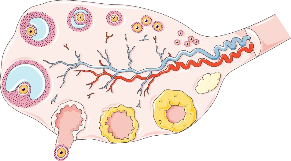
Image provided by Servier Medical Art (https://smart.servier.com), licensed under CC BY 4.0 (https://creativecommons.org/licenses/by/4.0/).
The Role of the Corpus Luteum in Pregnancy
If successful fertilization takes place, the life cycle of this temporary gland changes dramatically. The newly developing embryo quickly begins to secrete human chorionic gonadotropin (hcg), which is the exact marker tracked by home pregnancy tests. This vital hormone human chorionic gonadotropin acts as a rescue signal, instructing the ovary corpus luteum to override its scheduled breakdown and maintain high progesterone secretion. This steady hormonal support is what triggers the decidualization of the endometrium, locking the uterine lining in place to prevent menstruation and ensure a secure environment for implantation.
Beyond its primary role, the corpus luteum graviditatis – as it is known during gestation – also produces relaxin. This specialized hormone helps soften the maternal pelvic ligaments and aids in the structural relaxation of the uterus, while safely supporting the circulatory system during early pregnancy. The gland remains absolutely essential until around weeks 10 to 12, at which point the fully formed placenta takes over the responsibility of keeping progesterone levels elevated.
If the structure fails to supply enough progesterone before this transition occurs, a luteal phase defect can develop, significantly increasing the risk of early miscarriage. This is precisely why supplemental reproductive hormones are routinely prescribed to support patients throughout their first trimester in IVF treatments.
What Does the Corpus Luteum Look Like on Ultrasound?
On a transvaginal corpus luteum ultrasound, this structure typically appears as a small, round cyst-like shape with a thick, bright echogenic wall. It is most visible during the luteal phase of the menstrual month or throughout the first trimester of pregnancy.
When a scan reveals this structure in either the right or left ovary, it simply confirms that specific side successfully completed ovulation during that cycle. Doctors often identify it by switching to color Doppler imaging, which reveals a highly vascular “ring of fire” surrounding the tissue, confirming active corpus luteum function.

Corpus Luteum in the Right (or Left) Ovary: What Does It Mean?
Finding a corpus luteum in a specific ovary simply indicates the physical location of your most recent ovulation. There is absolutely no clinical significance to whether it forms on one side versus the other, as both ovaries are entirely capable of producing a healthy egg and supporting normal reproductive health.
On a scan, finding a corpus luteum in the right ovary means that the right side was responsible for releasing the egg during that particular menstrual cycle. While many women naturally alternate sides from one month to the next, it is completely normal for one ovary to ovulate more frequently than the other without impacting your overall fertility.
Corpus Luteum Cysts: When the Structure Doesn’t Break Down
Occasionally, rather than undergoing a normal regression, the gland fails to shrink and its internal cavity fills with fluid instead. When this happens, a corpus luteum cyst forms on the surface of the ovary. These specific fluid-filled sacs are incredibly common, almost always completely benign, and typically resolve entirely on their own over the course of a few weeks or menstrual cycles. In the vast majority of cases, these cysts produce absolutely no physical symptoms and are only discovered incidentally during a standard pelvic examination.
However, if the fluid accumulation continues or the sac grows unusually large, it can cause localized discomfort within the abdomen and pelvis. This fluid retention can sometimes mimic the initial signs of an ectopic pregnancy, making professional medical evaluation important. While a cyst is generally harmless, a large one can occasionally rupture or trigger a serious condition called ovarian torsion, where the ovary twists around its supporting tissues and cuts off its own blood supply.
Common Symptoms to Watch For:
- A dull, aching pelvic pain or localized pressure concentrated on one side.
- Persistent abdominal bloating or full sensation.
- Discomfort or deep pain experienced during sexual intercourse.
Most cysts require a simple approach of watchful waiting rather than surgery. However, you should see a doctor immediately if you experience sudden, severe pelvic pain, sharp abdominal distress accompanied by fever or vomiting, or unexpected heavy vaginal bleeding.
Corpus Luteum Defect and Fertility
A corpus luteum defect occurs when the temporary gland fails to produce adequate hormone levels, leading to what is clinically known as a luteal phase deficiency. When the function of the corpus luteum is compromised, the endometrium fails to thicken properly, making it incredibly difficult for a fertilized egg to successfully achieve implantation. This specific hormonal insufficiency can be triggered by chronic stress, thyroid dysfunction, or conditions like PCOS, often resulting in unexplained difficulty conceiving, a shortened menstrual cycle, or recurrent early pregnancy loss.
During stimulated IVF cycles, the protective corpus luteum development window is frequently disrupted by the medication protocols and the egg retrieval procedure itself. To safeguard the pregnancy, specialists routinely use targeted progesterone supplementation through injections, gels, or vaginal pessaries to ensure the uterine environment remains perfectly receptive.
Because IVF patients do not have to rely solely on how the luteum forms in the ovary, this medical support safely compensates for any underlying hormonal gaps. If you are exploring assisted reproduction and compiling an IVF FAQ and fertility questions checklist, understanding this protective protocol can offer immense peace of mind.
If you have questions about luteal phase support in IVF, the IVmed fertility team can help. Consult with the IVmed fertility experts today to learn more about personalized treatment protocols.
FAQ
The corpus luteum is a temporary endocrine gland that forms in the ovary just days after ovulation to produce progesterone. If you do not fertilise an egg during your cycle, the natural degeneration of the corpus luteum begins, and the tissue safely shrinks into a tiny scar called the corpus albicans.
The primary role of the corpus luteum in pregnancy is to secrete high levels of progesterone, which thickens the uterine lining to support embryo implantation. This vital structure remains active until the placenta completely takes over hormone production around weeks 10–12 of gestation.
On a pelvic scan, it appears as a small, round, thick-walled cystic structure on the ovulating ovary. When using color Doppler, ultrasound technicians can identify it by a distinct “ring of fire” flow pattern, which confirms active, healthy blood supply.
It simply confirms that your right ovary was the one that successfully released a mature egg during that specific menstrual cycle. Following ovulation, this structure turns into your primary hormone factory for the remainder of the month as the corpus luteum becomes essential for releasing progesterone to support potential conception.
A corpus luteum cyst is a common, benign functional sac that forms if the gland fails to break down after ovulation and instead fills with fluid. These temporary cysts are usually completely harmless, cause no pain, and resolve naturally within a few weeks.
Final Thoughts on the Corpus Luteum
Although the lifespan of corpora lutea is brief, this temporary gland plays a critical role in your monthly reproductive cycle. Understanding how it functions helps make sense of your fertility tracking, ultrasound results, and the essential role of the corpus luteum in pregnancy. Armed with this knowledge, you can better navigate your reproductive health and feel more confident during your medical checkups.
Have questions about your reproductive health or early pregnancy support? Contact the IVmed fertility team today to schedule a personalized consultation and get clear, expert answers.


For millions of individuals worldwide, navigating reproductive health comes with a silent, often misunderstood challenge. Chronic pelvic pain, grueling menstrual cycles, and unexplained exhaustion are

Understanding your ovarian reserve is one of the most vital steps when navigating family planning or preparing for assisted reproductive treatments. While blood tests measure

Ovulation typically happens around the middle of your menstrual cycle, opening a fertile window of a few days when the chance of getting pregnant is

A fraternal twin develops when a woman releases two separate eggs during ovulation, and each is fertilized by a different sperm. Also known as dizygotic,

When discussing male reproductive health, the focus typically falls on low sperm counts, but there is an opposite, much less talked-about phenomenon that leaves many

During your monthly cycle, you might notice sudden changes in your vaginal discharge. Among these variations, egg white discharge stands out as a distinct fluid

When navigating reproductive health, the true architecture of natural conception often relies entirely on a pair of sophisticated, yet highly vulnerable, biological pathways. Understanding where

When verifying a suspected pregnancy or monitoring early embryonic development, clinical precision is essential for proper medical management. While standard at-home kits offer basic screening,

We are pleased to announce that IVMED Family Agency will take part in the international fertility fair Wish for a Baby Köln 2025, held on

Join us TONIGHT, July 9th, at 8 PM CET, for a unique opportunity to hear directly from two top fertility experts as they dive into one of the hottest topics in reproductive medicine

It’s important for people to understand the connection between abortion and infertility because confusion can cause needless stress.

Fertility issues can be emotionally and physically draining, leaving many people unsure of what to do. We are here to support you because we recognize the profound impact these challenges can have on your life.